star facebook

MR Elastography now at Star Imaging, Bund Garden. MRE assesses the entire liver and detects fibrosis at an earlier stage than any other imaging method.

Medical Cases

April 2015

Star Imaging and Research Centre Medical case list

HISTORY

  • 128 Slice CT Scanner at Star and Imaging Research Centre
  • Cardiac Imaging Star and Imaging Research Centre
  • July-August Clinical Newsletter
  • Cranial Nerves Star and Imaging Research Centre

FINDING & CONCLUSION

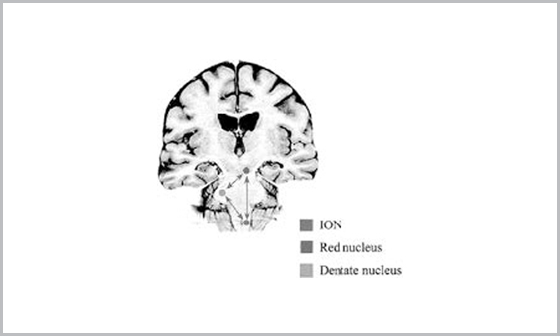
Small, well-defined T2 and flair hypointensity involving the left paramedian pons which shows blooming on the gradient images  with punctate enhancement on post contrast study–consistent with residual pontine cavernoma.Associated developmental venous anomaly noted with prominent veins coursing in the midline through the pons and across the left middle cerebellar peduncle. Left olivary nucleus of medulla shows hyperintense signal without significant swelling. No diffusion  restriction/presence of blood degradation products is seen. No e/o enhancement is noted.

Hypertrophic olivary degeneration – postoperative sequelae of pontine cavernoma resection

  • Hypertrophic olivary degeneration (HOD) is usually caused by a lesion in the triangle of Guillain and Mollaret and presents clinically as palatal tremor
  • The triangle consists of a set of connecting tracts including the cerebellar dentate nucleus, the brachium conjunctivum, and the contralateral oliva
  • The efferents from the dentate ascend through the superior cerebellar peduncle, cross in the decussation of the brachium conjunctivum inferior to the red nucleus, and then descend to the ION by way of the central tegmental tract. The triangle is completed by ION efferents crossing the midline, entering the inferior cerebellar peduncle, and terminating on the original dentate nucleu
  • Olivary deafferentation is thought to be the source of the ensuing hypertrophic degenerative changes. HOD is due to presumed transsynaptic degeneration resulting in vacuolation of neurons, an increase in the number of glial cells, demyelination, and shrunken neurons
  • Therefore, symptomatic palatal tremor and HOD are associated with lesions of the first two limbs of the triangle, but not with lesions involving olivodentate fibers
  • HOD usually occurs unilaterally and ipsilateral to the lesion if the lesion is in the brain stem or contralateral to the lesion if the lesion is in the cerebellum as identified on MR image
  • Three distinct MR stages in HOD are-
    • 1st stage – shows increased signal on T2-and proton density–weighted images without hypertrophy of the olive and occurs within the first 6 months of ictus
    • 2nd stage – both increased signal and hypertrophy and ends when hypertrophy resolves at approximately 3 to 4 years after ictus
    • 3rd stage – only shows increased signal and begins at the time hypertrophy resolves. This stage persists indefinitely