star facebook

Philips India arm partners with Star Imaging to start an MRI training school in the country! Read More

Service

128 slice CT

HIGHLIGHTS

With the installation of the premium 128 slice Ingenuity CT Scanner, we are one step closer to realising our mission of providing improved diagnostics, while minimising radiation exposure to the lowest possible levels.
Philips unique Adaptive Multicycle Reconstruction algorithm achieves temporal resolutions as fast as 53 milliseconds. This leads to excellent quality images with faster acquisition times.
These features give us a clear advantage when it comes to head-to-toe vascular system imaging, as well as in paediatric cardiac CT studies.
Moreover, the 128 slice CT scanner is also equipped with the award-winning iDose4 technology, ensuring that the patient is exposed to lowest possible X-rays, with up to 80% reduction in radiation dosage, while generating the best image quality.
  • Excellent image quality
  • Results in a more conclusive diagnosis
  • Faster image acquisition means less time spent in the scanner
  • Lowest possible radiation exposure (reduction by 80%)
  • Helps provide accurate diagnosis in paediatric imaging cases
  • CT scans, especially neurovascular CTA of the head and neck, are non-invasive techniques that reduces procedure time and minimises patient discomfort
  • 3D CT combined with CT angiography helps provide critical information to plan surgical procedures in the event of kidney diseases
  • Yields images with better temporal and spatial resolution, greater anatomic coverage of the patient, more consistent enhancement with lesser volume of intravascular contrast material and high quality 2D reformation and 3D reconstruction owing to acquisition of an isotropic data set
  • Equipped with the award-winning iDose4 technology, ensuring that the patient is exposed to lowest possible X-rays, with up to 80% reduction in radiation dosage
  • Provides non-invasive 3D anatomy in post endovascular/surgical repair assessment and successfully evaluates all associated complications within and outside the vascular lumen
  • 128 slice MDCT scanner offers better diagnostic interpretation and display of the entire aorta non-invasively and at lowest radiation dose (iDose)
  • In case of kidney diseases, provides pre-operative interactive display of a 3D model of the affected kidney and its vascular supply to the surgeons
  • Advent of MDCTA challenges old gold standard of DSA in quantifying atherosclerotic disease load in patients with PAD
  • Provides accurate depiction of complex cardiovascular anatomic features both before and after surgery in cases of paediatric congenital heart disease
  • Provides high quality, three dimensional image data sets to study cerebro-vascular anatomy
  • Provides 3D anatomy of grafts, is non-invasive and successfully visualises the grafts, anatomosis and distal vessel
CT Scan in Pune Diagnostic Radiology Clinic in Pune

One of the key diagnostic elements guiding the therapeutic approach in the acute phase of an ischemic stroke is the presence or absence of intracranial large vessel occlusion (LVO). This can be detected using neurovascular CTA of the head and neck. CTA, being a non-invasive technique saves both procedure time and patient discomfort, as compared with DSA and MRA. It provides high quality, three dimensional image data sets to study cerebro-vascular anatomy.

In staging renal cell carcinoma, the goal of any imaging study is to identify patients who have a resectable tumor and can be cured by means of surgical intervention. Three dimensional CT combined with CT angiography has the potential to provide all the critical information needed to characterise the lesion and plan the surgical procedure.
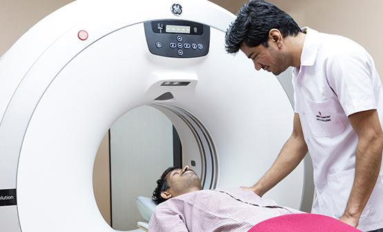
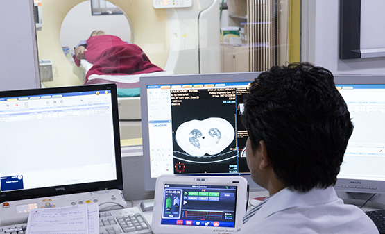
 Our 128 slice CT scanner provides pre-operative interactive display of a 3D model of the affected kidney and its vascular supply to the surgeons.

Previously, DSA was the only established reliable imaging technique to quantify atherosclerotic disease load in patients with PAD. Advent of MDCTA now challenges this old gold standard, along with other non-invasive techniques such as magnetic resonance angiography (MRA).

Our 128 slice MDCT scanner offers better diagnostic interpretation and eloquent anatomical and pathological display of the entire arterial tree, non-invasively and at lowest radiation dose (iDose) optimizing maximum arterial enhancement, without venous contamination is a challenge in assessing distal run off of symptomatic limb.

CT angiography is important in the evaluation of pediatric congenital heart disease. It can be used for accurate depiction of complex cardiovascular anatomic features both before and after surgery. Evaluation of post treatment complications can also be performed.

CT facilitates assessment of extracardiac systemic and pulmonary arterial and venous structures.

Compared with the previous CT scan machines, 128 slice scanner yields images with better temporal and spatial resolution, greater anatomic coverage of the patient, more consistent enhancement with lesser volume of intravascular contrast material and high quality 2D reformation and 3D reconstruction owing to acquisition of an isotropic data set. These are achieved with an additional advantage of reduced radiation dose to patient. CT can be considered the first line imaging technique for evaluation of suspected vascular ring or sling, mapping of the aortopulmonary collateral arteries in patients with severe right ventricular outflow tract obstruction and evaluation of an anomalous pulmonary and systemic venous return.

Cardiac coronary angiography study allows ostium evaluation, presence/absence of vascular remodelling, percentage stenosis, characterisation of the plaque, evaluation of its extent and site, involvement of branch origin and evaluation of distal vessel. Limitations include very high calcium deposition within the vessel walls obscuring the lumen due to blooming artifacts. It has a negative predictive value in the range of 98% for ruling out significant in stent restenosis. Assessment of bypass grafts and distal runoffs by invasive coronary angiography is cumbersome and often requires extra procedure time, contrast load, and radiation exposure. Cardiac CT provides 3D anatomy of grafts, is non-invasive and successfully visualises the grafts, anastomosis and distal vessel.

contact details

Please fill in details

Address: Deccan

Joshi Hospital Campus, Opposite Kamla Nehru Park,
Erandawane, Pune 411004

Address: Bund Garden

Connaught Place, Ground floor,
Bund Garden Road, Pune - 411 001

Address: Akluj

Shamrao Patil Medical Complex,
Akluj - 413 101

Address: Wakad

Solitaire Business Hub, Opp. Ambience hotel,
Vishnu Dev Nagar, Wakad,
Pimpri-Chinchwad 411057

CALL US NOW

FOR YOUR INFORMATION

The machine takes your pictures, just like a camera. The advanced machines give far better quality which helps the radiologist and your doctor understand your problem.

No. You just have to lie down for 15-20 minutes or for as long as required for the study.

The required time depends upon what study needs to performed, cooperation from the patient, and requirement of sedation or contrast.

It depends on what kind of scan is advised to you. Our front office will help you with these queries.

Yes, you will get a CD for the scan. You will get the films in fifteen minutes

Study of the scan images require lot of dedicated time and expertise of radiologist. We try to report studies in the best possible time.

You can pay in cash or by debit/credit card. Star does not charge extra when patients pay with debit/credit cards.