For Enquiries and Appointments call +91 97136 11611 (Pune Centres) | Akluj: 02185-224444
ABOUT STAR IMAGING
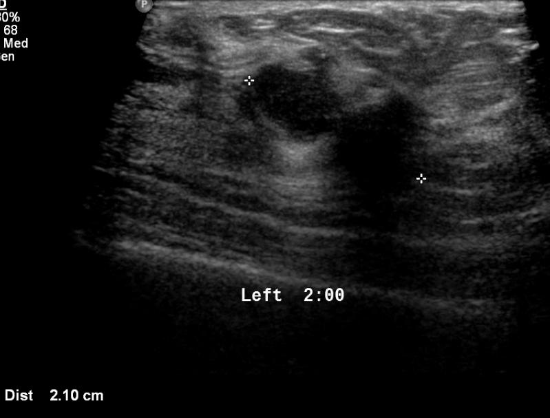
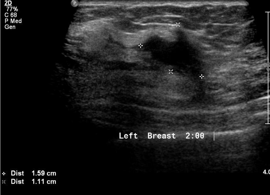
Services
Divisions
Patients Hub
Doctor hub
Our locations
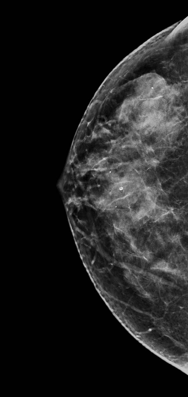
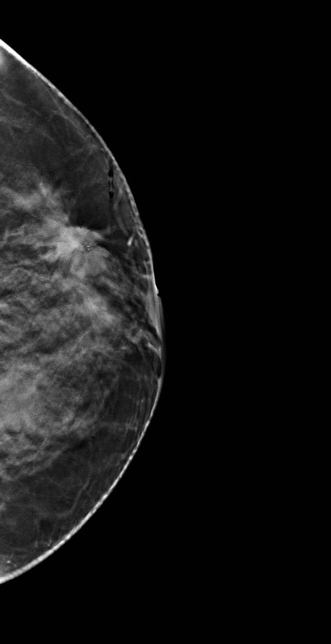
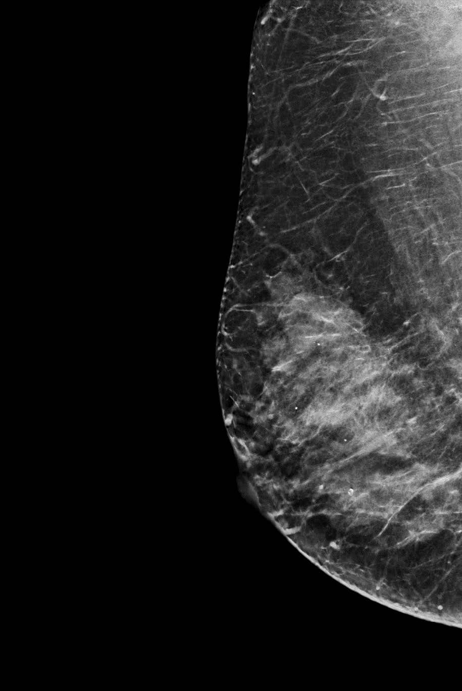
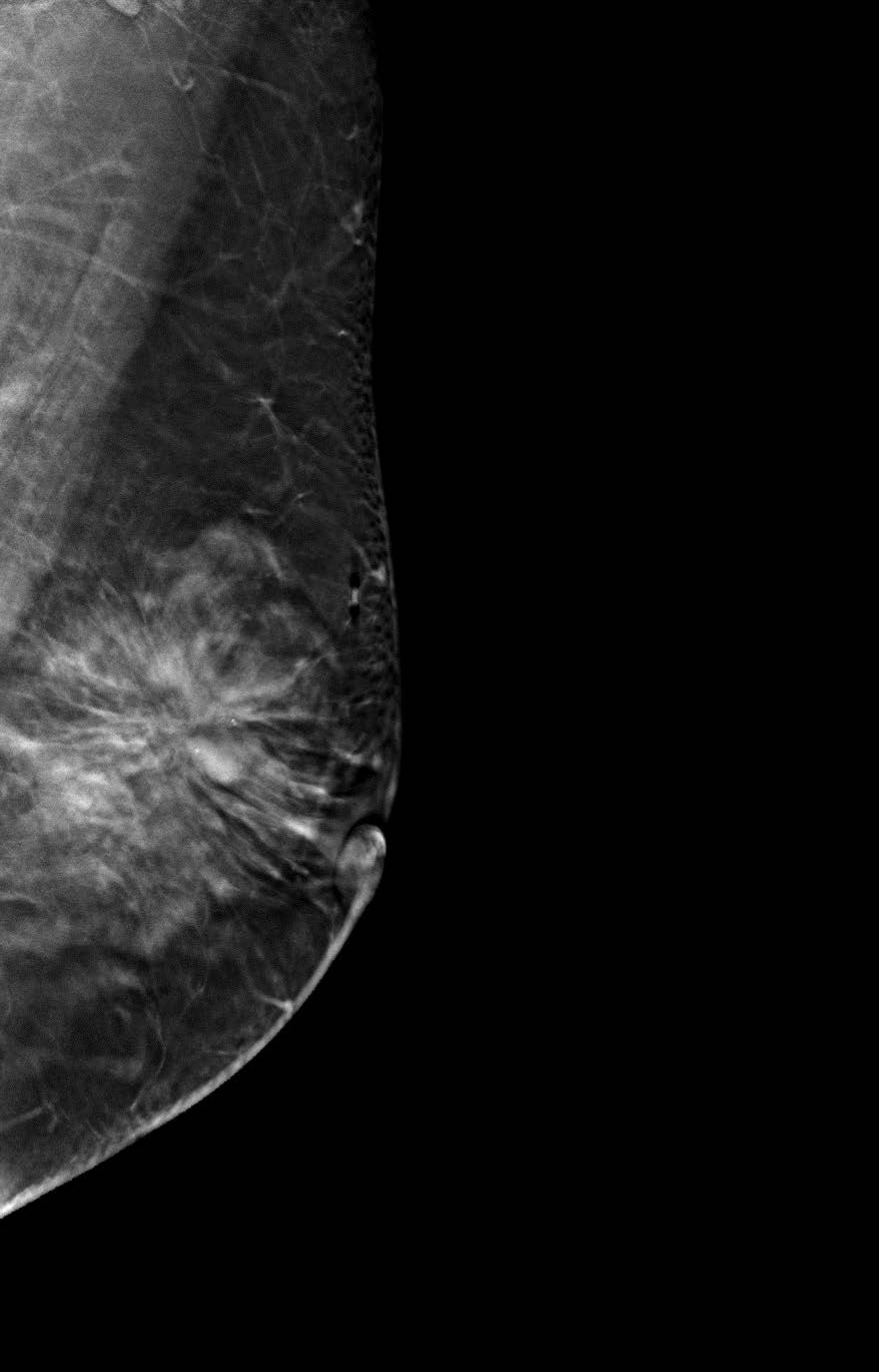
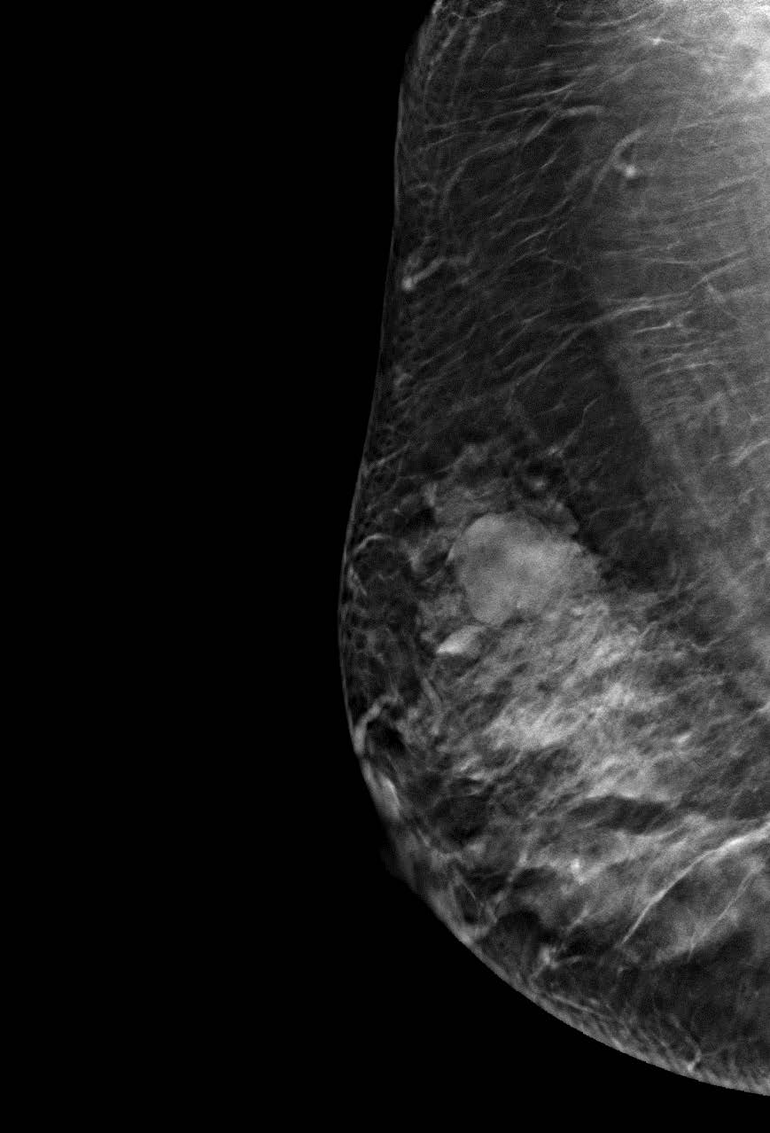
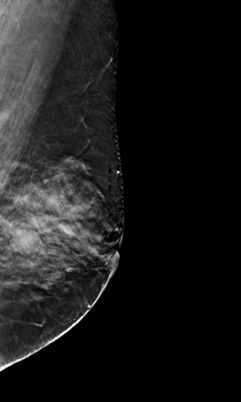
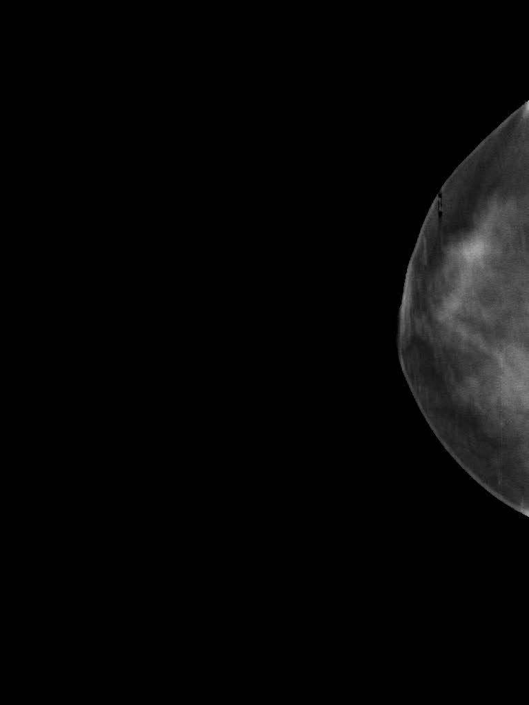
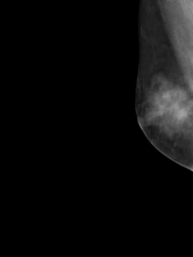
Digital Tomosynthesis