Tear of the anteroinferior labrum with paralabral cyst
Tear of the entire superior, posterosuperior, posterior and posteroinferior labrum with large posteroinferior para-labral cyst extending into the axillary region
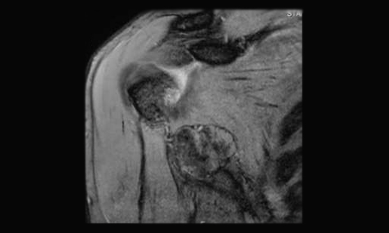
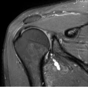
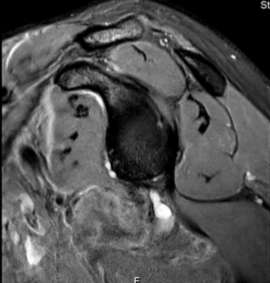
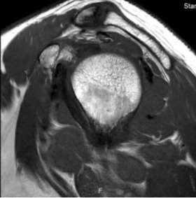
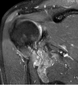
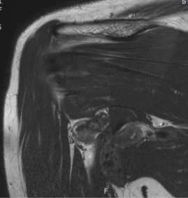
Well-defined, large, lobulated soft tissue lesion seen in the right axilla partly insinuating into the axillary pouch. Lesion is mildly hyperintense with dark areas within it on T2, which bloom on the gradient echo images (due to hemosiderin staining). It is isointense on T1 and enhances heterogeneously
PVNS represents a benign, hypertrophic proliferative disorder of the synovium characterized by villous, nodular, and villonodular proliferation and pigmentation from hemosiderin. It may affect the joints, bursae, or tendon sheaths. It can appear in either a diffuse or, less commonly, a focal form within the joint
The knee is the most frequently involved joint, followed by the hip, ankle, and shoulder
Conventional radiographs of joints affected by PVNS may appear normal or may demonstrate periarticular soft-tissue swelling. Joint spaces and bone mineralization are characteristically preserved until late in the disease. Bone erosions are common in joints with a tight capsule, such as the hip and ankle. Extrinsic erosion, often with well-defined sclerotic margins, of the underlying bone is the most common osseous abnormality
Computed tomography depicts these lesions as either diffuse thickening of the tissue about the joint or as a localized soft-tissue mass. The attenuation of the lesion may be somewhat increased relative to that of muscle owing to the presence of hemosiderin
MR imaging reveals characteristic features of a heterogeneous, diffuse, synovially based, plaquelike thickening that often is associated with nodularity. Associated joint effusion is common, particularly in large joints such as the knee, but the effusion is generally surrounded by thickened synovial rinds of hemosiderin-laden tissue. The signal intensity of the synovial thickening is intermediate to low on T1-weighted images. Low signal intensity also predominates on T2-weighted MR images, owing to the preferential shortening of T2 relaxation time caused by hemosiderin, an effect that is accentuated at higher field strength. This effect is particularly pronounced on gradient-echo images, which demonstrate an enlargement of the low-signal-intensity areas (“blooming”) that is caused by magnetic susceptibility artifact. The blooming effect, which specifically signifies the presence of hemosiderin as the cause of low signal intensity, is nearly pathognomonic of PVNS at MR imaging
Malignant PVNS is rare and difficult to distinguish, both pathologically and radiologically, from multiple local recurrences of benign disease unless there is metastatic involvement of the lungs or lymph nodes
Rheumatoid arthritis may show similar significant synovial proliferation. Associated findings of uniform reduction in joint space and subchondral geodes
Synovial hemangioma and hemophiliac arthropathy may show similar MR imaging findings (caused by repetitive intraarticular hemorrhage and synovial hemosiderin deposition), diffuse intraarticular PVNS can be distinguished from these conditions because it is not associated with either serpentine vascular channels (hemangioma) or a clinical history of hemophilia