star facebook

To book an appointment: Wakad: +917942611128 | Deccan: +917942611126 | Bund Garden: +917942611127 | Akluj: 02185-224444 | Baner: +917942611129

Medical Cases

September 2014

Dr. Varshaa Hardas, Dr. Sanjay Desai

Star Imaging and Research Centre Medical case list

HISTORY

  • 67 year old lady with a history of a lump in the right breast
  • No other clinical complaints
  • FNAC done with results positive for IDC
  • Outside Mammography: DBP, not very informative
  • Sent for Digital Tomosynthesis and USG Abdomen/Pelvis to assess liver

FINDING & CONCLUSION

  • Mammography revealed a irregular solid lesion in the right breast upper quadrant with mild architectural distortion
  • Mild spiculations also noted
  • No foci of abnormal microcalcifications noted
  • Few foci of scattered benign macrocalcifications noted
  • In drawing of the right nipple noted, suggesting retroareolar extension of the disease
  • Right axillary nodes noted
  • Vascular calcifications noted bilaterally
  • Modeartely dense breast parenchyma bilaterally
  • No abnormal lesion in the left breast
  • BI-RADS VI–RIGHT BREAST
  • Large solid lesion in the right periareolar region extending from 12 to 2 o’ clock, with irregular margin and some internal vascularity
  • Extension in the retroareolar region also noted
  • Lesion measures 2.6 x 2.3 x 1.3cm
  • Small BI-RADS III lesion in left breast noted of 7mm at 10 o’ clock position. Strain value suggest benign nature
  • Elastography strain values is 5, also suggestive of malignant neoplastic etiology
  • Heterogeneous liver echotexture with no focal lesions
  • Large solid lesion in the cervix suspicious for neoplastic etiology
  • This lesion had moderate vascularity
  • Enlarged heterogeneous uterus with a focal echogenic bright leiomyoma in the fundus
  • Ovaries not well seen. Endometrium was normal
  • Multiple Para-aortic lymphadenopathy was noted. Largest was 2.8cm
  • No ascites
  • Large solid lesion in the cervix with moderate vascularity of 3.7 x 2.8cm size
  • Enlarged bulky heterogenous uterus with focal intramural bright echogenic lipo-leiomyoma in the fundus of 3.2 x 2.5cm size
  • Ovaries were not well seen
  • Normal endometrium with indentation from the leiomyoma
  • Based on these usg findings furthur evaluation with CT abdomen and pelvis was advised
  • Biopsy of the cervical lesion was also suggested
  • Slightly bulky uterus showing heterogeneous enhancing mass lesion involving the uterine cervix-likely to be neoplastic
  • A well-defined lesion in relation to (most likely within) the left side of the uterine fundus with soft tissue and fat could represent a lipoleiomyoma
  • Multiple enlarged retroperitoneal (from the level of superior mesenteric artery upto the aortic bifurcation), bilateral common & internal iliac and retrocrural lymph nodes
  • Patient underwent TRU_CUT core biopsy of breast and cervical lesion
  • Breast – Invasive ductal carcinoma grade II
  • Carvix – Adenocarcinoma

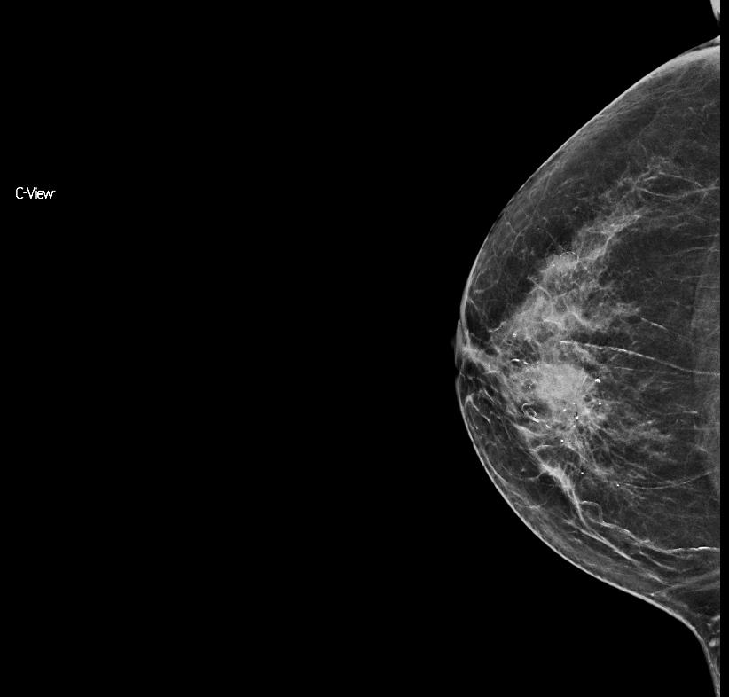
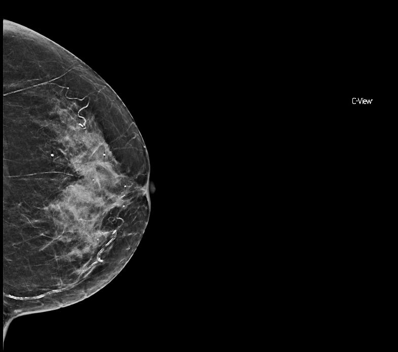
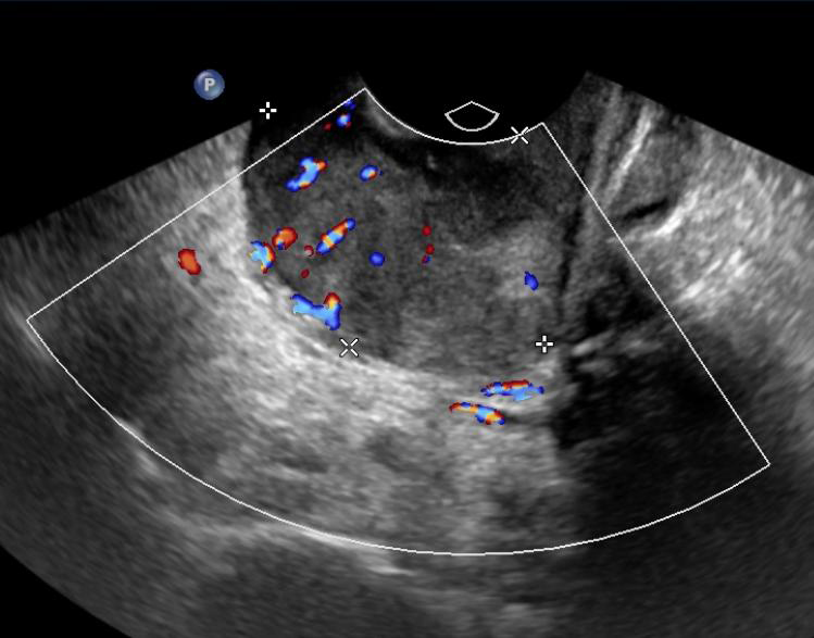
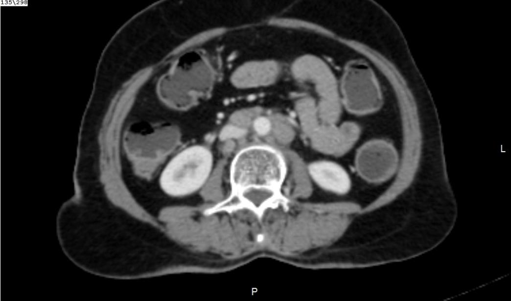
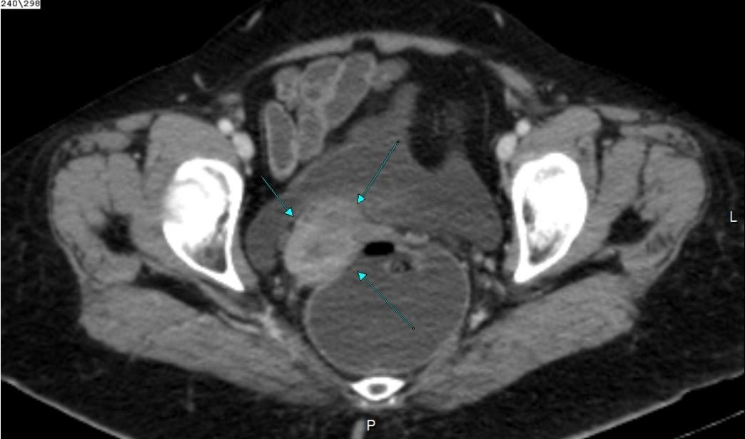
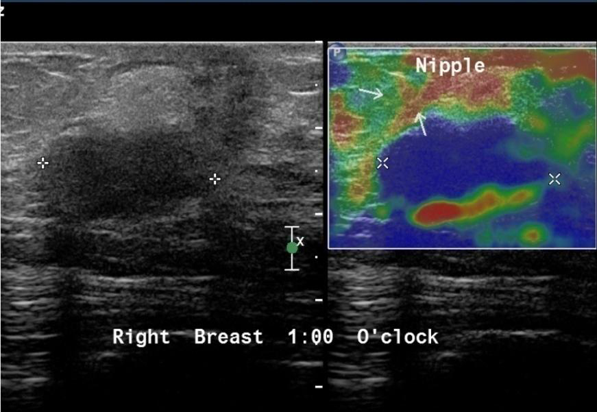
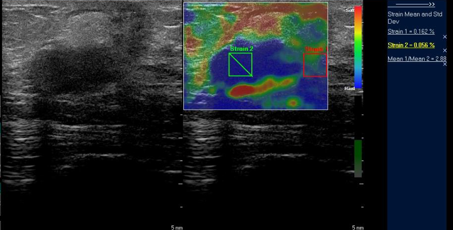
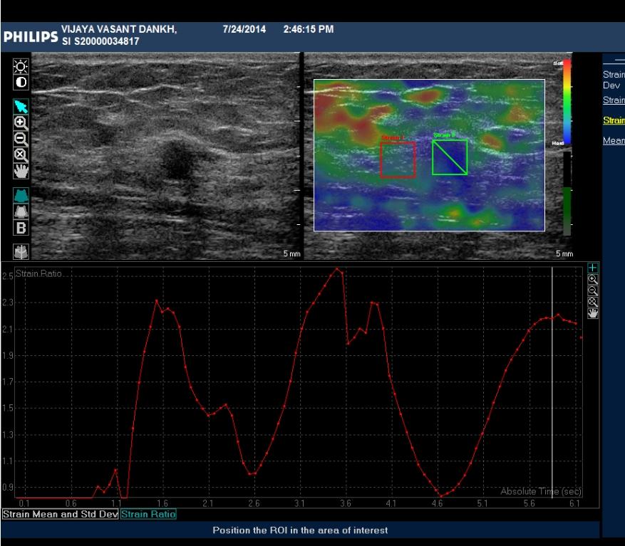
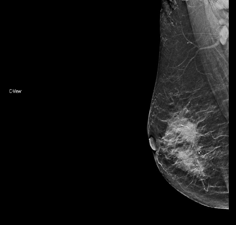
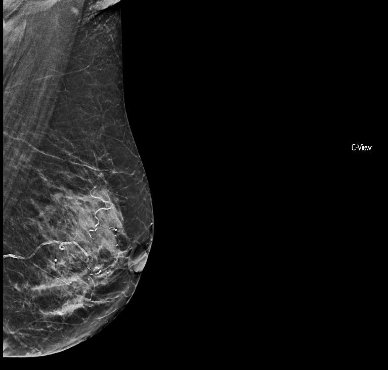
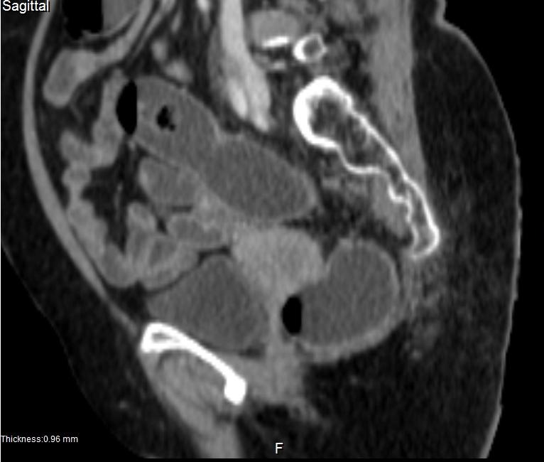
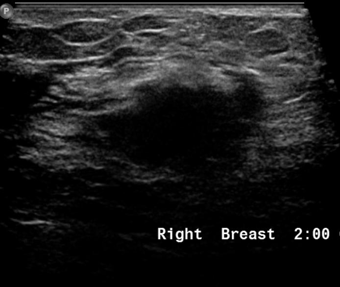
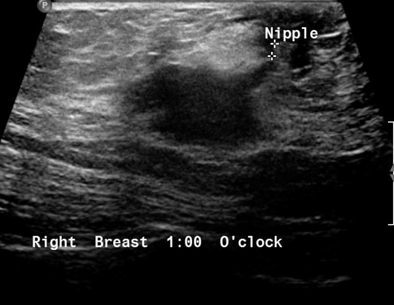
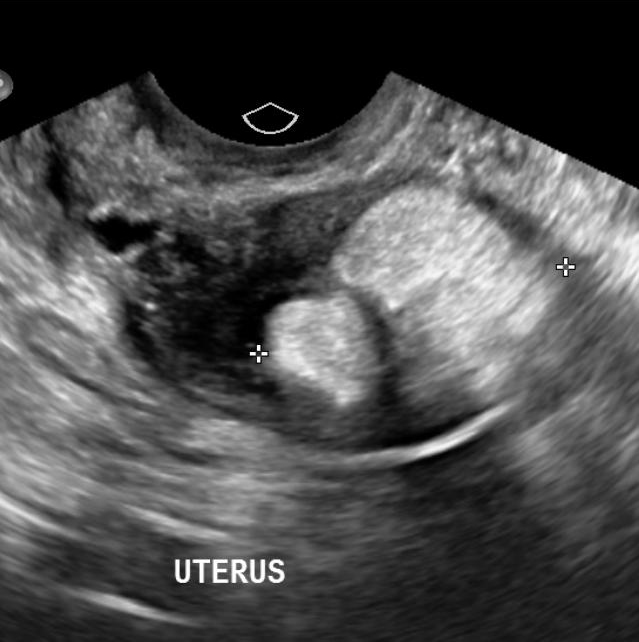
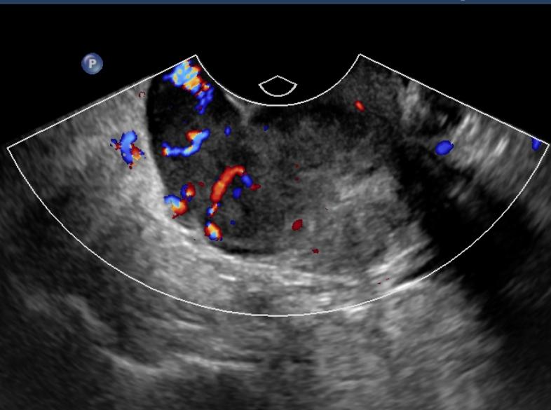
SYNCHRONOUS DUAL PRIMARY MALIGNANCIES–BREAST & CERVIX

  • Breast and Ovarian synchronous carcinomas are known to occur
  • The synchronous occurrence of carcinoma of the uterine cervix and the breast is a rarity
  • No definite link has been established between the two
  • OCP use increases the incidence of both cancers but not necessarily together
  • Occurrence of Lipo-Leiomyoma is also rare

SYNCHRONOUS DUAL PRIMARY MALIGNANCIES –BREAST & CERVIX

  • AGARWAL RASHI. Synchronous dual malignancy: Successfully treated cases. Journal of Cancer Research and Therapeutics, Vol. 3, No. 3, July-September, 2007, pp. 153-156
  • Verstovsek S et al. Synchronous primary cancers of the breast and cervix: planning multidisciplinary primary treatment [clinico-pathological conference]. Am J Clin Oncol.2000 Feb;23(1):99-103
  • Sharma DN. Synchronous occurrence of carcinoma of the uterine cervix and of the breast . European Journal of Surgical Oncology Volume 25, Issue 5, Pages547–548, October 1999 
  • Prieto A, Crespo C, Pardo A et-al. Uterine lipoleiomyomas: US and CT findings. Abdom Imaging. 25 (6): 655-7
  • Tsushima Y, Kita T, Yamamoto K. Uterine lipoleiomyoma: MRI, CT and ultrasonographic findings. Br J Radiol. 1997;70 (838): 1068-70