star facebook

MR Elastography now at Star Imaging, Bund Garden. MRE assesses the entire liver and detects fibrosis at an earlier stage than any other imaging method.

Medical Cases

October 2014

Star Imaging and Research Centre Medical case list

HISTORY

  • 3 year male
  • c/o left arm pain
  • Palpable bony tenderness
  • Normal range of movements
  • No fever
  • H/o trivial trauma (fall)

FINDING & CONCLUSION

  • Irregular area of altered marrow signal intensity in diaphysis of middle one third of humerus
  • It appears slightly hyperintense on T2 and STIR images and hypointense on T1 w images
  • Heterogeneous moderately intense post contrast enhancement is noted
  • The lesion shows a thin irregular sclerotic margin and multiple internal septae. There is diffuse adjacent marrow edema. No fluid levels are apparent
  • CT images reveals irregular predominantly cortical lesion encroaching on medullary cavity. Thinning of the lateral cortex is noted with cortical breaks at multiple places. Associated circumferential periosteal reaction is noted
  • Intense post contrast enhancement is noted in this region. Para-osseous soft tissue component is also evident with mild post contrast enhancement extending into the adjacent muscles and intermuscular fat planes. Mild bowing of the humerus with convexity towards left side
  • Ewing’s Sarcoma  –

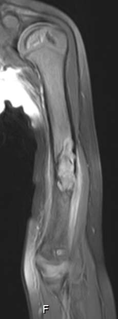
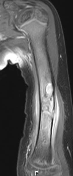
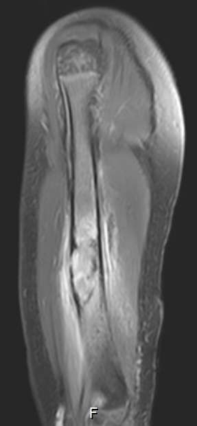
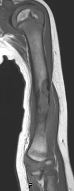
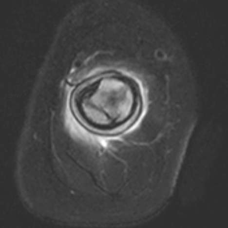
    Considering MRI findings-
    Irregular area of altered marrow signal intensity in diaphyseal location with cortical break and associated periosteal thickening and adjacent paraosseous soft tissue.

  • Osteofibrous dysplasia/ossifying fibroma-

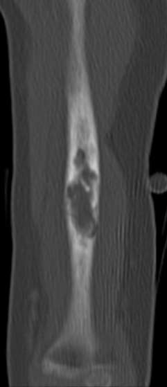
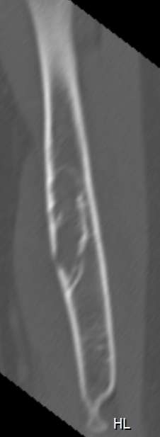
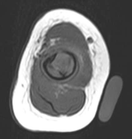
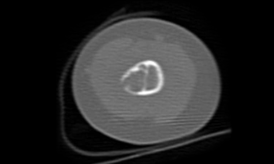
    Considering CT findings –
    Cortical and diaphyseal location of lesion.
    Bowing of shaft of humerus.
    Peripheral sclerotic margin.

  • Histopathological Diagnosis
  • Osteofibrous Dysplasia
  • Also called ossifying fibroma
  • Osteofibrous dysplasia (OFD) is a rare, benign, self-limited fibro osseous lesion that is seen almost exclusively in the diaphysis of the tibia
  • Other long bones are also involved but rarely
  • OFD is seen in patients aged < 20 years
  • The lesion generally involves the anterior cortex and may cause bowing of shaft of long bones
  • OFD typically manifests as an intracortical lytic lesion, which is generally well marginated and is often surrounded by a zone of sclerosis
  • Multiple lucencies may be present within the cortex, with intervening sclerotic areas
  • The involved cortex may be expanded and/or thickened
  • Periosteal reaction is rare, but when present, it is thick, solid, and chronic–appearing
  • OFD seldom progresses radiographically during childhood, and progression halts when the patient reaches skeletal maturity
  • While reporting any bone tumour/lesion consider CT also with MRI because sometimes MRI will show different findings
  • Considering all imaging findings we should take account of history also (as in our case pt is having h/o trivial trauma. So the soft tissue in the MRI could be secondary to pathological fracture in lesion)
  • Sometimes typical lesions can occur at atypical site (as in our case humerus is rare site for Osteofibrous dysplasia)
  • Park YK, Unni KK, McLeod RA, Pritchard DJ: Osteofibrous dysplasia: Clinicopathologic study of 80 cases. Hum Pathol 1993;24(12):1339-1347
  • Kahn LB: Adamantinoma, osteofibrous dysplasia and differentiated adamantinoma. Skeletal Radiol 2003;32(5):245-258
  • Papagelopoulos PJ, Mavrogenis AF, Galanis EC, Savvidou OD, Inwards CY, Sim FH: Clinicopathological features, diagnosis, and treatment of adamantinoma of the long bones.Orthopedics 2007;30(3):211-215
  • Levine SM, Lambiase RE, Perchprapa CN: Cortical lesion of the tibia: characteristic appearance at conventional radiography. Radiographics 2003;23 (1): 157 -177